Conny Waters – AncientPages.com – Research from the University of Gothenburg has revealed significant health issues among Sweden’s Viking Age population, including severe oral and maxillofacial diseases, sinus and ear infections, and osteoarthritis.

Credit: Pixabay – Kyraxys – Public Domain
This conclusion comes from a study where Viking skulls were analyzed using advanced X-ray techniques. Previously, research focused on examining teeth from Varnhem’s Viking Age population in Västergötland—a site known for its numerous ancient graves and well-preserved skeletons. Building on this work, odontologists have now employed modern computed tomography (CT) scans to study entire skulls.
Findings published in the British Dental Journal Open indicate that the fifteen individuals examined exhibited a wide range of diseases. The CT scans revealed pathological bone growths in the cranium and jawbone, indicating various infections and conditions.
Several individuals exhibited sinus or ear infections, leaving detectable traces in the surrounding bone structures. Additionally, evidence of osteoarthritis and various dental diseases was observed. The skulls examined belonged to adults who passed away between the ages of 20 and 60.
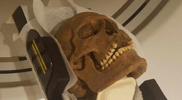
The skulls of Viking-era individuals were examined with modern computed tomography, in the search for infections, inflammations and other diseases. Credit: Carolina Bertilsson
The study was led by Carolina Bertilsson, an assistant researcher at the University of Gothenburg and a dentist with Sweden’s Public Dental Service. It involved collaboration with specialists in dental radiology from the University of Gothenburg and an archaeologist from Västergötlands Museum. Together, they conducted examinations and analyzed images using CT scans. These scans provide three-dimensional images that allow researchers to examine skeletal damage in detail, layer by layer, across different parts of the skull.
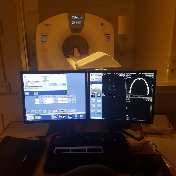
Computed tomography provide 3D photos and the possibility of advanced image analysis where layer by layer of bones, jaw bones and teeth are studied in detail. Credit: Carolina Bertilsson
“There was much to look at. We found many signs of disease in these individuals. Exactly why we don’t know. While we can’t study the damage in the soft tissue because it’s no longer there, we can see the traces left in the skeletal structures,” says Carolina Bertilsson in a press release, and continues:
“The results of the study provide greater understanding of these people’s health and wellbeing. Everyone knows what it’s like to have pain somewhere, you can get quite desperate for help. But back then, they didn’t have the medical and dental care we do, or the kind of pain relief – and antibiotics – we now have. If you developed an infection, it could stick around for a long time.”
See also: More Archaeology News
The study is described as a pilot study. One important aspect was to test CT as a method for future and more extensive studies.
“Very many of today’s archaeological methods are invasive, with the need to remove bone or other tissue for analysis. This way, we can keep the remains completely intact yet still extract a great deal of information,” says Carolina Bertilsson.
Written by Conny Waters – AncientPages.com Staff Writer